Why Most Horse Tendon Injuries Come Back and How to Stop It
Last Updated::
One bad step is all it takes. A horse can go from full fitness to months of box rest because of a single tendon injury. If you’ve ever felt the back of your horse’s leg go hot and puffy after a workout, you know how quickly the worry sets in. How bad is it? What’s the best horse tendon recovery treatment? And how long before they’re back to work?
Tendon injuries are one of the most common reasons sport horses go lame or retire early. Somewhere between 11 and 30% of racing Thoroughbreds will deal with one during their career. But science has come a long way. Treatments that were still experimental ten years ago now have real clinical trial data behind them, and better imaging tools mean vets can spot damage much earlier, before a small strain turns into a serious tear.
This guide walks you through everything in order. You’ll learn what equine tendons actually are and why they get hurt, how to spot the warning signs early, what’s happening inside the tendon as it heals, which treatments have the strongest evidence behind them, and what it takes to keep your horse sound after recovery.
What Is a Tendon Injury in Horses?
Tendons are tough, rope-like bands of tissue that connect muscle to bone. In the horse’s lower leg, two tendons run down the back of the cannon bone between the knee and the hoof: the superficial digital flexor tendon (SDFT) and the deep digital flexor tendon (DDFT). The suspensory ligament sits alongside them, providing extra support.
A tendon injury occurs when these fibres are stretched beyond their limit and begin to tear. The damage can range from a mild strain (a few fibres disrupted) to a complete rupture. Because tendons have a limited blood supply compared to muscle or skin, they heal slowly and the repaired tissue is never quite as strong as the original.
The SDFT is by far the most commonly injured tendon in performance horses, accounting for 75 to 93% of all equine tendon injuries. The mid-cannon bone region of the front legs is affected in 97 to 99% of cases. The DDFT, which runs deeper and attaches inside the hoof, is injured less often but is harder to diagnose and treat because of its hidden location.
Why the Superficial Digital Flexor Tendon (SDFT) Is the Most Commonly Injured Tendon
The SDFT works like an elastic band. During galloping and jumping, it stretches to store energy and then snaps back to help propel the horse forward. This makes it highly efficient, but it also means the tendon operates dangerously close to its breaking point during every fast workout.
A 2021 review by O’Brien and colleagues at the Royal Veterinary College explained that during high-speed galloping, the equine SDFT typically stretches by 10 to 16%, while laboratory tests show the tendon ruptures at roughly 12 to 21% stretch. That leaves a margin of just a few percentage points between normal use and catastrophic failure. The review explained that the real problem is not a single overload but the gradual build-up of tiny, invisible tears (microdamage) over weeks and months of training. Each workout chips away at the tendon’s structure, and as the horse ages, its ability to repair that everyday damage declines. This is why tendon injuries tend to appear in older, seasoned horses rather than young ones, and why the same review found that 23 to 67% of horses treated with rest alone re-injure within two years.
How to Spot a Tendon Injury Early
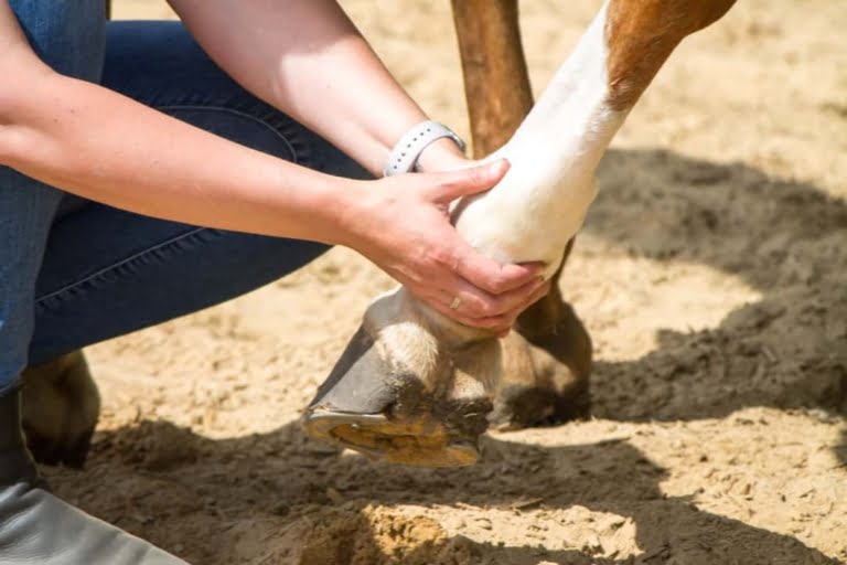
Catching a tendon injury early can shorten the recovery time for a horse tendon injury by months. There are four signs to watch for.
- Heat is usually the first warning. Run your hand down the back of each cannon bone. If one leg feels warmer than the other, that local temperature rise points to inflammation underneath.
- Swelling follows shortly after and, in SDFT injuries, shows up as a thickening or puffiness along the back of the leg.
- Lameness ranges from a subtle unevenness at the trot to obvious reluctance to bear weight in severe cases.
- Pain on touch confirms the location and gives your vet an initial sense of how serious the damage is.
Your veterinarian will use ultrasound to see inside the tendon. This shows the size and location of any damaged area (called a core lesion), the percentage of the tendon’s width that is affected, and whether the fibres are still organised or disrupted. The size of the lesion on ultrasound is the single strongest predictor of outcome.
For injuries inside the hoof, particularly deep digital flexor tendon tears, MRI is essential because ultrasound cannot penetrate the hoof wall. A 2025 review in the Equine Veterinary Journal by Scharf and colleagues compared different MRI systems for equine foot imaging and highlighted that serial MRI scans are now considered important for monitoring recovery and deciding when to increase exercise.
Healing of Tendons
Unlike bone, which can rebuild itself to nearly original strength, a damaged tendon heals with scar tissue that is weaker and less elastic than the original. A review in the Annual Review of Biomedical Engineering by Voleti and colleagues described four overlapping stages that every tendon injury goes through.
Phase 1: Inflammation (Days 1 to 10). A blood clot forms at the injury site. The body sends in clean-up cells that clear away damaged fibres and release chemical signals to start the repair process. This is when heat, swelling, and pain are at their worst. Complete rest is critical.
Phase 2: Repair (Weeks 2 to 6). Specialised repair cells called fibroblasts move in and begin laying down new collagen to bridge the gap between torn fibres. The problem is that this early collagen (type III) is thinner and weaker than the collagen in healthy tendon (type I). A study by Williams and colleagues in Research in Veterinary Science measured this difference directly: normal equine tendon is made almost entirely of type I collagen, while scar tissue contains 20 to 30% of the weaker type III. New blood vessels also grow into the area during this phase.
Phase 3: Remodelling (Months 2 to 6). The weaker type III collagen is gradually swapped for stronger type I, and the fibres start lining up along the direction of pull. Controlled, gradual exercise becomes important here because gentle loading tells the repair cells which direction to lay the new fibres. But be warned: the horse may look and feel sound during this phase even though the tissue is still fragile inside. Apparent comfort is not the same as structural readiness.
Phase 4: Maturation (6 to 18 Months). The collagen fibres strengthen through cross-linking, and the tissue slowly gains stiffness. Even after full maturation, the repaired area typically remains weaker than the original tendon, which is why re-injury rates are high and why long-term management matters.
The four phases of equine tendon healing, from acute inflammation to long-term maturation.
Treatment Options for Horse Tendon Injuries
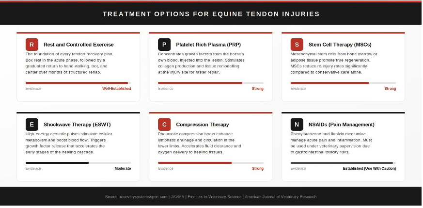
Treatment has come a long way from rest and bandaging. Today’s best recovery plans combine several approaches, each targeting a different part of the healing process. Here are the six main options, in the order they are typically introduced.
1. Rest and Controlled Exercise
Every horse tendon recovery treatment starts with rest. In the first 10 days (the inflammatory phase), box rest takes all weight-bearing stress off the damaged fibres. As healing progresses, your vet will design a step-by-step exercise programme: hand-walking first (usually 10 to 20 minutes, once or twice daily), then slow trot work, then small paddock turnout, and eventually canter — spread over many months. Each step up happens only when an ultrasound scan confirms that the tissue inside is ready. Total recovery time typically ranges from 8 to 16 months depending on how severe the injury is.
2. Anti-Inflammatory Medication (NSAIDs)
Drugs like phenylbutazone (“bute”) and flunixin meglumine are the two most commonly prescribed NSAIDs in equine medicine and are regularly used in the first days after a tendon injury to manage pain and reduce inflammation. They provide important comfort during the acute phase, but they must be used carefully. A review in The Veterinary Journal by Lees and Toutain found that phenylbutazone is the most likely to cause gut problems, followed by flunixin, then ketoprofen. The most serious risk is a condition called right dorsal colitis — an inflammation of part of the large intestine that can cause colic, diarrhoea, weight loss, and fluid build-up under the belly. A newer drug called firocoxib has a lower gut-risk profile and may be a better option for horses that need longer-term pain relief. Your vet will choose the right drug, dose, and duration for your horse.
3. Platelet Rich Plasma (PRP) Therapy
PRP therapy takes a sample of your horse’s own blood, spins it in a machine to concentrate the platelets and their growth factors, and then injects that concentrate directly into the injured tendon. The growth factors stimulate the repair cells to produce more collagen and reorganise the healing tissue.
A controlled study in the Journal of Orthopaedic Research by Bosch and colleagues found that PRP-treated tendons had significantly more collagen, better fibre organisation, and improved strength after 23 weeks compared to untreated controls. A 2016 trial in BMC Veterinary Research by Geburek and colleagues tested PRP in 20 horses with naturally occurring SDFT injuries and found that the PRP group showed less lameness earlier (by week 8 versus week 12 in the control group) and more organised repair tissue on imaging.
One important note: a 2021 critical review in Frontiers in Veterinary Science pointed out that while PRP clearly improves tissue quality in the short term, high-quality evidence that it prevents re-injury over the long term is still limited. The 2024 analysis in JAVMA found only one study that reported PRP-specific re-injury data. So PRP’s main value right now is in helping the tendon heal better and faster in the early stages, rather than as a proven shield against future breakdown.
4. Stem Cell Therapy (MSCs)
Stem cell therapy is the treatment option with the strongest evidence for reducing re-injury in horse tendon injuries. Mesenchymal stem cells (MSCs) — usually taken from your horse’s own bone marrow or fat tissue — are injected into the damaged area, where they encourage the growth of better-quality repair tissue instead of plain scar.
The clinical results are compelling. Pacini and colleagues reported in 2007 that 9 of 11 racehorses treated with bone-marrow stem cells returned to racing within 9 to 12 months and were still racing without re-injury at 2 years. The conventionally treated control group had a 100% re-injury rate within 12 months. A much larger study by Godwin and colleagues in 2012 followed 141 client-owned racehorses and found a re-injury rate of 27.4% with stem cell treatment, compared to 53 to 56% in historical controls. Most recently, a 2023 comparison of 213 Thoroughbreds by Salz and colleagues found that horses receiving bone-marrow stem cells were significantly more likely to return to racing and complete five or more races.
The big-picture view comes from the 2024 JAVMA systematic review and meta-analysis by M’Cloud and colleagues, which pooled data from 21 studies (17 in the statistical analysis). The combined odds ratio for re-injury with stem cells or stem-cells-plus-PRP versus conservative treatment was 0.29 — meaning a 71% reduction in re-injury odds. That is a meaningful clinical improvement.
A word of honesty. The same review flagged that study quality across the field is poor: only 2 of 21 studies were rated good quality, none blinded the examining vet or owner, and several landmark results used historical controls rather than randomised comparisons. Stem cells clearly reduce re-injury risk, but they are not a guarantee of full athletic recovery. The best results are seen in younger horses, with smaller lesions, treated sooner after injury with adequate cell numbers.
5. Shockwave Therapy (ESWT)
Shockwave therapy sends focused pulses of sound energy into the damaged tendon. This stimulates the tendon’s own repair cells (called tenocytes) to increase their activity, promotes new blood vessel growth, and triggers the release of growth factors. A 2007 study in the Equine Veterinary Journal by Bosch and colleagues at Utrecht University confirmed these metabolic effects in ponies, and a follow-up study in 2009 showed that focused shockwaves affected collagen structure and gene activity in treated tendons.
Shockwave therapy becomes especially interesting when combined with PRP. A 2017 study in Frontiers in Veterinary Science by Seabaugh and colleagues found that applying shockwaves to PRP samples in the laboratory increased one key growth factor (TGF-B1) by up to 46% and another (PDGF-BB) by up to 219%. While this is laboratory evidence rather than a clinical trial in living horses, it provides a strong scientific reason for combining the two therapies.
6. Compression Therapy
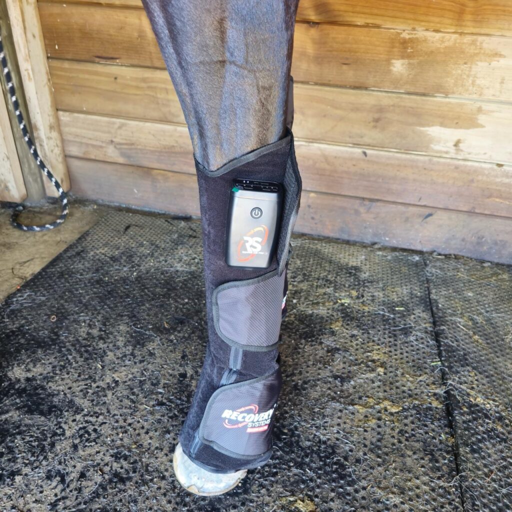
Intermittent pneumatic compression — using inflatable boots that squeeze and release in a rhythmic cycle — is well established in human sports medicine and is now gaining ground in equine practice. The boots push fluid through the lymphatic vessels and veins, helping to deliver fresh, oxygenated blood to healing tissue and flush out inflammatory waste. Horses are especially prone to lower-limb swelling because they have almost no muscle below the knee to pump fluid back up on their own, making external compression support particularly useful during tendon recovery.
A 2023 study in the American Journal of Veterinary Research by Koch and Schnabel at North Carolina State University tested compression therapy on six Thoroughbreds using a tracking method that follows fluid movement through the lymphatic system. They found that pneumatic compression led to significantly faster fluid flow to key drainage points in the foreleg compared to untreated controls. The study supports the use of compression therapy as a helpful addition during tendon recovery, where better circulation means more nutrients reaching the healing tissue and faster removal of swelling.
Veterinary practitioners working with performance horses in Asia have observed these benefits in practice. Dr Dan Shaw, a veterinarian who works with horses at the Turf Club, has spoken about applying human compression therapy principles to equine recovery. He points out that in human sports medicine, research consistently shows the more effort put in during the post injury period, the better the outcomes, and he sees the same logic carrying over to horses. He’s also said he’s a believer in anything that reduces inflammation and improves blood flow, and that Recovery Systems Equine products deliver on both.
Dr Sukumar Shetty, a veterinarian at the Singapore Polo Club, has observed that polo horses frequently sustain tendon and ligament injuries from running at high speed, hard stops, sharp turns, and contact during play. He describes intermittent compression as a strong recovery concept because it brings more blood circulation to the limbs, which means more oxygen and nutrition reaching the tissues. In his view, that’s what speeds up healing, especially in tendon and ligament cases, and warming the legs while boosting circulation to the lower limbs helps horses feel and move better.
Support Your Horse’s Tendon Recovery with Targeted Compression
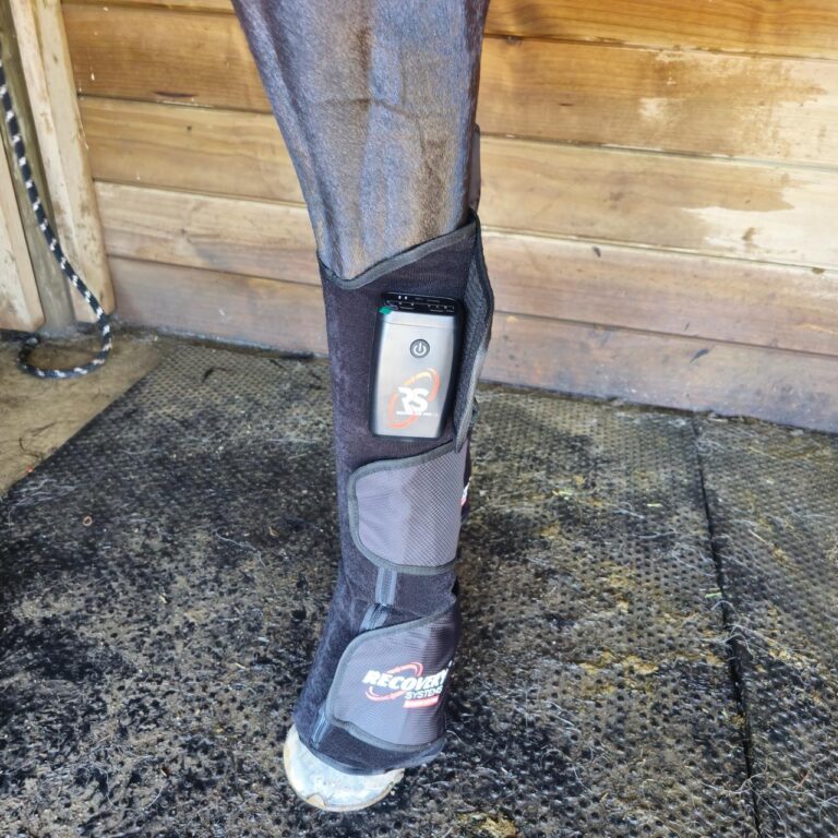
The Recovery Systems Equine Air Pro delivers intermittent pneumatic compression across all four limbs, supporting lymphatic flow and circulatory health during every phase of tendon rehabilitation.
Treatment options for equine tendon injuries
How to Prevent Re-Injury After Recovery
A successful tendon recovery does not end when the horse returns to work. The healed tissue is structurally different from the original, and the risk of re-injury stays elevated — particularly in the first 12 months after return to full exercise. A sustainable prevention plan includes the following steps.
- Keep up regular imaging. Even after the horse is back in full work, periodic ultrasound (or UTC where available) lets your vet catch early signs of trouble before the horse shows any outward symptoms. UTC is especially useful here because it can detect fibre problems that look completely normal on standard ultrasound.
- Get the footing right. Excessively deep, soft ground and rock-hard surfaces both increase tendon strain. Consistent, well-maintained footing reduces the mechanical load on every stride.
- Warm up properly. Cold tendons are stiffer and more vulnerable to microdamage. A gradual warm-up that builds speed and intensity progressively prepares the tendon for the elastic demands of galloping and jumping.
- Continue compression and cool-down support. Compression therapy, cold hosing, and structured cool-down routines after exercise help manage the low-level inflammation that accompanies training.
- Do not rush. The most common cause of re-injury is returning to high-intensity work too quickly. Follow your vet’s graduated exercise plan even when the horse feels perfectly sound, because the tissue inside may not be ready.
- Know when to change the goal. If the original lesion occupied more than 50% of the tendon’s width, the horse is over 10 years old, the injury is a repeat, or serial imaging shows no improvement, the most responsible approach may be a shift to a lower-impact discipline or retirement from competition. That is not failure — it is evidence-based care that puts the horse’s welfare first.
Conclusion
Horse tendon recovery treatment has moved well beyond rest and hope. The science now supports a step-by-step approach: prompt diagnosis with ultrasound or MRI, responsible use of anti-inflammatory medication, regenerative therapies like PRP and stem cells to improve tissue quality and reduce re-injury risk, compression therapy to support circulation, and a structured, imaging-guided rehabilitation programme that advances only when the tissue inside the tendon is genuinely ready.
Every tendon injury is different. Severity, location, the horse’s age, and their athletic demands all shape the recovery path. What the evidence consistently shows is that a proactive, science-guided plan, one that starts with your vet and continues through every phase of healing, gives horses the best chance of returning to meaningful work.
Frequently Asked Questions
Can a Horse Be Ridden After a Tendon Injury?
Many horses do return to ridden work after a tendon injury, but the timeline depends on how severe the damage is. Mild strains may allow a gradual return to light riding within 3 to 6 months, while a more seriously injured tendon can require 12 to 18 months before it is safe to carry a rider. Your vet will use serial ultrasound scans to check whether the tissue inside has reorganised enough to handle the load. Rushing this step is the single biggest reason horses re-injure. The 2023 Equine Veterinary Journal study of 213 Thoroughbreds showed that horses following a structured 12-month rehabilitation programme with stem cells had the best chance of sustaining a long term racing career.
Can a Horse Fully Recover From a Tendon Injury?
Full recovery to the original tissue quality is uncommon. Tendons heal with scar tissue that contains 20 to 30% of the weaker type III collagen instead of rebuilding the original type I structure, according to a study by Williams and colleagues in Research in Veterinary Science. That scar tissue is never quite as strong or elastic as the original. However, many horses achieve a functional recovery that lets them return to their previous level of work. The 2024 JAVMA meta-analysis by M’Cloud and colleagues found that stem cell treatment reduced re-injury odds by 71%, which is the strongest current evidence for long term recovery quality. Younger horses with smaller lesions treated earlier tend to have the best outcomes.
Can a Horse Recover From a Bowed Tendon?
Yes, many horses with bowed tendons return to work. A bowed tendon is a visible, curved swelling along the back of the cannon bone caused by fibre damage in the SDFT. Under conservative management (rest and controlled exercise only), roughly 50 to 60% of racehorses with moderate to severe bows return to training, and the numbers are higher for sport and pleasure horses that do not compete at speed. Bowed tendon horse treatment follows the same principles as any SDFT injury. Mild bows respond well to rest, cold therapy, corrective shoeing, NSAIDs, and compression therapy. Moderate to severe cases benefit from adding platelet rich plasma or stem cell injections, guided by ultrasound. Regenerative therapies, particularly stem cells, improve these numbers significantly.
Can a Horse Recover From a Deep Digital Flexor Tendon Tear?
How well a horse recovers from a deep digital flexor tendon tear comes down to exactly where the damage sits. A 2021 study in Frontiers in Veterinary Science used standing low-field MRI to assess horses with digital flexor tendon sheath swelling and found that when the injury sat near the pastern bone, at the point where the tendon changes shape, the results were poor. Only 3 out of 13 horses (23%) made it back to their previous level of work. Horses with navicular bursa involvement fared slightly better, with around 35% returning to full use after medical treatment. On the other end of the scale, a 2022 study by Hewitt-Dedman and colleagues looked at injuries right at the tendon’s attachment point on the coffin bone and found a much more encouraging picture, with about 67% of those horses getting back to the work they were doing before. MRI is essential for these cases because standard ultrasound cannot image deep digital flexor tendon lesions inside the hoof.
How Do You Treat a Superficial Flexor Tendon Injury in Horses?
Treatment of a superficial digital flexor tendon injury starts with rest and anti-inflammatory medication in the acute phase, followed by a vet-guided exercise programme that builds up slowly over months. Advanced options include platelet rich plasma (which speeds tissue healing, as shown in the 2016 BMC Veterinary Research trial by Geburek and colleagues), stem cell therapy (which reduces re-injury by 71% according to the 2024 JAVMA analysis), shockwave therapy (which stimulates repair cells), and compression therapy (which supports blood flow and swelling reduction). Superficial digital flexor tendon horse recovery typically takes 8 to 16 months depending on severity, with each step guided by ultrasound imaging. The 2023 study of 213 Thoroughbreds by Salz and colleagues confirmed that horses receiving bone-marrow stem cells alongside a 12-month rehabilitation programme had the best chance of returning to racing.
How Do You Treat a Torn Tendon in a Horse?
Treating a torn tendon in a horse follows a step-by-step approach. In the first 10 days, strict box rest and NSAIDs (such as phenylbutazone or flunixin meglumine, the two most commonly prescribed equine NSAIDs) manage pain and control inflammation. From there, the healing process depends on a graduated exercise programme designed by your vet, starting with hand-walking and progressing through trot work to canter over many months. For moderate to severe tears, regenerative therapies like platelet rich plasma and mesenchymal stem cells are injected directly into the injured tendon to promote better tissue repair rather than plain scar tissue formation. Compression therapy supports the healing process by boosting circulation in the lower limbs. The 2024 JAVMA meta-analysis confirmed that combining stem cells with structured rehabilitation gives the best long term outcomes.
How Long Does a Deep Digital Flexor Tendon Injury Take to Heal?
Deep digital flexor tendon injuries generally take longer than SDFT injuries, often 9 to 18 months or more. Because the deep digital flexor tendon sits in a mechanically complex area inside the hoof, the healing process is slower and harder to monitor. Your vet may recommend MRI at intervals to track progress, as ultrasound cannot provide enough detail for lesions inside the foot. The 2025 Equine Veterinary Journal review by Scharf and colleagues highlights that serial MRI is increasingly considered standard for monitoring deep digital flexor tendon recovery and deciding when to increase exercise loading.
How Long Does a Tendon Injury Take to Heal in Horses?
The horse tendon injury recovery time depends on severity. The inflammatory phase lasts about 10 days, the repair phase continues through approximately week 6, remodelling runs from months 2 to 6, and final maturation takes 6 to 18 months, as described in the Annual Review of Biomedical Engineering by Voleti and colleagues. In practical terms, mild strains may reach functional recovery in 3 to 6 months, moderate injuries take 8 to 12 months, and severe injuries need 12 to 18 months or more. Each step should be guided by ultrasound imaging rather than by how comfortable the horse appears, because an injured tendon that seems healed on the outside may still be fragile inside.
How Long Does It Take a Horse to Recover From a Superficial Digital Flexor Tendon Injury?
Recovery from a superficial digital flexor tendon injury follows the same four-phase healing process as any tendon injury, but the timeline depends on severity and treatment. Mild SDFT strains may allow a return to work within 3 to 6 months. Moderate core lesions typically need 8 to 12 months of imaging-guided rehabilitation. Severe injuries with significant cross-sectional involvement can take 12 to 18 months or longer. The 2021 review by O’Brien and colleagues found that 23 to 67% of horses managed with rest alone re-injure within two years, which is why most equine vets now recommend combining structured exercise with at least one regenerative therapy. The 2012 study by Godwin and colleagues followed 141 racehorses treated with stem cells and found a re-injury rate of 27.4%, compared to 53 to 56% with conventional care.
What Are the Treatment Options for a Bowed Tendon in a Horse?
Treatment options for a bowed tendon range from conservative to regenerative. Conservative care includes rest, controlled exercise, cold therapy, corrective shoeing, NSAIDs, and compression therapy. Regenerative options include platelet rich plasma, stem cell injections, and shockwave therapy. In rare cases of complete rupture, surgery may be discussed. The right combination depends on the severity shown on ultrasound, with the size of the core lesion relative to the total tendon width being the key decision-making factor. Most equine vets now recommend at least one regenerative therapy alongside structured rehabilitation for moderate to severe bowed tendon injuries. Under conservative management alone, roughly 50 to 60% of racehorses return to training. Adding stem cells improves those odds significantly, as confirmed by the 2024 JAVMA meta-analysis showing a 71% reduction in re-injury.
What Can I Do to Help My Horse Recover From a Tendon Injury?
Work closely with your vet and follow the exercise plan exactly, advancing only when ultrasound or UTC imaging confirms the tissue is ready. Do not rely on how the horse looks or moves, because the injured tendon may feel sound while the tissue inside is still fragile. Keep up regular check-ups to monitor the healing process. Provide well-maintained footing for turnout and exercise. Consider pneumatic compression boots to support circulation in the lower limbs, where horses have almost no muscle to pump fluid on their own. Make sure nutrition and hoof care are on point. And if the original lesion was large (more than 50% of the tendon’s width), have an honest conversation with your vet early about whether a return to high-speed work is realistic, or whether a shift to a lower-impact discipline is in the horse’s best interest. Contact your equine vet for a personalised recovery plan.
What Causes Bowed Tendons in Horses?
Bowed tendons happen when the SDFT is stretched beyond its limit and the fibres tear. The main causes are overexertion during training or racing, fatigue (which reduces the tendon’s ability to absorb strain), hard or uneven ground, poor leg shape (long sloping or upright pasterns, long toe/low heel), and jumping into fast work without warming up. Age-related wear and tear also reduces how much strain the tendon can handle. The 2021 Equine Veterinary Journal review by O’Brien and colleagues explained that the real driver is cumulative microdamage that builds up over weeks and months of training, gradually weakening the tendon until it fails. Bandage bows caused by over-tight wrapping are a separate issue and involve the tissue around the tendon rather than the tendon itself.
What Does a Torn Tendon Look Like in a Horse?
On the outside, a torn tendon shows up as swelling along the back of the cannon bone, warmth to the touch, and usually lameness. In bowed tendons, the swelling creates a visible outward curve in the tendon profile. On ultrasound, the vet will see a dark area (called a hypoechoic core lesion) inside the tendon where the fibres are damaged. The size of that dark area as a percentage of the total tendon width is the key number your vet uses to grade how serious the injury is and to plan treatment. A large retrospective study of 469 Thoroughbreds in Hong Kong found that a core lesion below 50% of the tendon’s width gave a 29 to 35% chance of racing again, while lesions above 50% dropped to just 11 to 16%.
What Is the Fastest Way to Heal a Tendon Injury?
There is no shortcut. Tendon healing follows a biological timeline that cannot be safely rushed. The fastest evidence-based approach is to combine early vet diagnosis with a multi-pronged treatment plan. Platelet rich plasma improves tissue quality and speeds early-phase repair (Bosch et al., 2010). Stem cells cut re-injury odds by 71% (M’Cloud et al., 2024). Compression therapy supports blood flow and helps clear swelling from the lower limbs (Koch et al., 2023). These therapies work within the biological healing process, not around it. The biggest gains come from early detection and disciplined, imaging-guided rehabilitation.
What Is the Most Common Tendon Injury in Horses?
The superficial digital flexor tendon injury is by far the most common, accounting for 75 to 93% of all equine tendon injuries. The 2021 Equine Veterinary Journal review by O’Brien and colleagues confirmed that the SDFT’s energy-storing function and the gradual build-up of microdamage during fast work make it especially vulnerable. The front legs are affected in nearly all cases, with the mid-cannon bone region involved in 97 to 99% of injuries. The deep digital flexor tendon and the suspensory ligament are injured less frequently, but tendons and ligaments in the lower limb share a limited blood supply, which is why all of these structures heal slowly. The flexor tendons (both superficial and deep digital flexor) bear the greatest mechanical load during galloping and jumping, which explains why they are the most injury-prone.
What Is the Difference Between Tendon and Ligament Injuries in Horses?
Tendons connect muscle to bone, while ligaments connect bone to bone. In the horse’s lower leg, the flexor tendons (the superficial digital flexor tendon and the deep digital flexor tendon) and the suspensory ligament all run down the back of the cannon bone and work together to support the fetlock joint during movement. Injuries to tendons and ligaments share many of the same causes, including overexertion, poor footing, and fatigue. The healing process is also similar for both: slow, scar-based repair that never fully matches the original tissue. The key difference is that suspensory ligament injuries are sometimes harder to detect on standard ultrasound and can involve the branches of the ligament near the fetlock, which adds complexity to diagnosis. Treatment principles for both tendon injuries and ligament injuries follow the same evidence-based approach of rest, controlled exercise, regenerative therapies, and long term imaging-guided rehabilitation.
| WRITTEN BY Michael Lyons Michael Lyons is a recovery specialist with three decades of tech and Meditech experience and 15,000+ hours in endurance sports coaching. The author is not a licensed physician or medical professional. The health information in this article represents the author’s research and personal perspective, not professional medical advice. MEDICALLY REVIEWED BY Ayman Shafique, PharmD Ayman Shafique is a licensed pharmacist with expertise in pharmacology and medical writing. Her work focuses on reviewing and developing evidence-based health and wellness content, ensuring scientific accuracy, clarity, and alignment with current pharmaceutical research. She specialises in translating complex pharmacological and medical information into accessible, reliable content for public and professional audiences. |
| Medical Disclaimer This article is for informational and educational purposes only and should not be considered medical advice. The author is a recovery specialist, not a licensed medical doctor or healthcare provider. Health-related claims discussed in this article are based on emerging research and should not replace professional medical consultation. Many claims remain theoretical and require further scientific validation. Individual results may vary, and these concepts are not widely accepted as standard medical practice. Before using any PEMF therapy device or making health decisions based on this article, please consult a qualified healthcare professional — especially if you have pre-existing medical conditions, are pregnant, have implanted medical devices such as pacemakers, or are taking medications. Commercial Disclosure This website sells PEMF therapy products, including BioMat technology. The author and company have a commercial interest in promoting these products. If you are experiencing a mental health crisis or suicidal thoughts, contact your local emergency number immediately. In the United States, call or text 988. Internationally, see the International Association for Suicide Prevention directory. |
Originally Published::
Author:
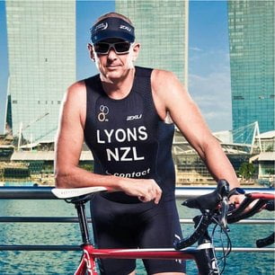
Michael Lyons is a biohacking and recovery specialist with three decades of tech and Meditech experience and 15,000+ hours in endurance sports coaching.